|
3DVisA Resources
3DVisA Index of 3D Projects: Digital Arts
Tomography Art by Kai-hung Fung
Kai-hung Fung is a medical doctor and a specialist in diagnostic radiology at Pamela Youde Nethersole Eastern Hospital, Hong Kong SAR, China.
He also applies radiology to visual arts. Using his experience of 3D Computer Tomography (CT) for medical diagnosis, he creates artistic images of the human body based on data acquired from CT scanners.
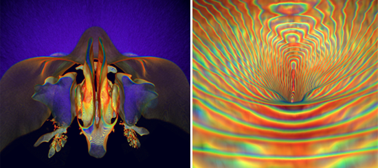
Fig. 1. Kai-hung Fung, What lies behind our nose?, Computed Tomography (CT) imaging, 2007. Fig. 2. Maternal connection. 3D CT of the human umbilicus rendered with rainbow technique, 2007. © Kai-hung Fung. Reproduced with kind permission.
Dr Kai-hung Fung is a winner of the 5th annual International Science and Engineering Visualization Challenge 2007,
sponsored by Science, the journal of the American Association for the Advancement of Science (AAAS), and the National Science Foundation (NSF). His entry, entitled What lies behind our nose?, has been awarded the first prize (tie) in the photography category. It was created
using 3D computed tomography (CT) of the human nose and paranasal sinuses, and a special rendering method developed by Dr Kai-hung Fung, known as 'rainbow technique'. He is also interested in developing stereoscopic artwork using 3D CT.
The Rainbow Technique in 3D Computed Tomography
by Kai-hung Fung
The 'rainbow technique' is a new method of art representation using contour lines to define 3D space or object, each contour line being in the form of a colourful rainbow. Similar to pointillism, this new concept of colour representation vitalize the artwork transforming it into one that sparkle with colours. Due to its complexity, it is only made possible with modern computer technology.
My interest in using 3D computed tomography (CT) as a tool for creative art originated just a few years ago, perhaps around 2003 when we acquired a Toshiba 16-slice multi-detector row CT in our hospital for medical imaging. With this new line of CT scanner, high resolution reconstructed 3D images can be obtained due to the improved performance of the CT scanner (thinner slices, faster scanning time) coupled with availability of more powerful computer workstation and more sophisticated software for 3D reconstruction. We used Vitrea®2 software by Vital Images, Inc.
Although most people are quite satisfied with the preset range of colour rendering provided by the 3D software (usually involving a limited choice of colours), I tried to explore how I can utilize the full colour spectrum in my 3D rendering. With perseverance and a lot of luck, I was able to realise the concept of the 'rainbow technique' and produce 3D CT artworks successfully with this technique in 2005.
In fact 3D image in CT is formed by staking together hundreds of very thin slices (about 0.5 to 1 mm in thickness). Stepping artefacts are inherent in this staking process particularly along sloping edges. The stepping artefacts will show up as contour lines in the 3D object only at certain critical settings.
The CT image in fact is a plot of the CT values in small picture elements called voxels. The usual rendering method is to apply a grey-scale to these values to form the image. The grey-scale can be manipulated by shifting the window level and window width in CT just like changing the gamma and latitude in a film. The manipulation could help to show different structures (e.g. air, fat, soft tissue or bone) with different CT values.
In colour 3D rendering, different colour spectra are applied instead of a grey scale. Before applying the colour, an algorithm in the form of a curve can be applied to bias the colour rendering towards specific CT range.
So by adjusting the window level and window width together with the use of a very narrow band width algorithm, a critical level could be reached for structures showing specific CT values so that the surface of the 3D structure starts to break up into contour lines. Applying a rainbow spectrum at this point produces the 'rainbow technique'.
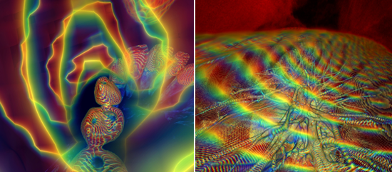
Fig. 3. Kai-hung Fung, Rotting teeth. 3D CT of the lower set of human teeth rendered with rainbow technique, 2007. Fig. 4. Lung surface. 3D CT of the human lungs rendered with rainbow technique, 2007. © Kai-hung Fung. Reproduced with kind permission.
The rainbow effect is particularly noticeable in strongly curved surface and at close-up. It adds to the 3D perception by showing up as contour lines instead of smooth surface with gradation. Furthermore it acts like a shutter blind instead of a curtain in separating 3D space. Structures behind can be clearly seen instead of blurred. Those structures that have CT values outside the critical range will be rendered in the usual way, giving additional room for artistic creativity. The rainbow effect can also be perceived stereoscopically in virtual reality if stereoscopic pair of images is obtained and viewed with appropriate tools such as a stereoscope.
Of course, apart from the 'rainbow technique' there are many ways in rendering the 3D CT e.g. using semi-transparency to produce artistic effect. However, in my opinion the 'rainbow technique' is particularly appealing aesthetically.
Project dates: Ongoing since 2005.
Sources and further details:
Kai-hung Fung (2006),
The rainbow technique: an innovative approach to the artistic presentation of 3D computed tomography, Leonardo, vol. 39, No. 2, pp. 101-103, color plate A.
Kai-hung Fung (2006), 'Artwork Using 3D Computed Tomography: Extending Radiology into the Realm of Visual Art', Leonardo, vol.39, No. 3, pp.187-191.
Lester, B. (2007), Science and Engineering Visualization Challenge 2007, Special feature,
Science, 28 September, vol. 317. no. 5846, pp. 1858 - 1863; DOI: 10.1126/science.317.5846.1858 (includes a slide show of the winning entries).
Record originally compiled by Anna Bentkowska-Kafel, 11 September 2006. Co-authored by Dr Kai-hung Fung, 24 October 2007. Last updated: 31 October 2007.
3DVisA gratefully acknowledges the help of Dr Kai-hung Fung with preparation of this record.
© Kai-hung Fung and 3DVisA, 2006.
Back to the list of 3D projects
|